 breast.oncosurgery@gmail.com
 +91 9871413426
 Home
 Testimonials
 Blogs
 Contact



Mammogram Says Normal but You Still Have a Lump
- Blog
- Mammogram Says Normal but You Still Have a Lump
You felt something unusual in your breast. Naturally, you scheduled a mammogram. The report came back normal.
But there is one problem: you can still feel the lump.
This situation can be confusing and frightening. Many women wonder, “If my mammogram is normal, why can I still feel a lump?” or “Does a normal mammogram mean I can safely ignore it?”
The short answer is no.
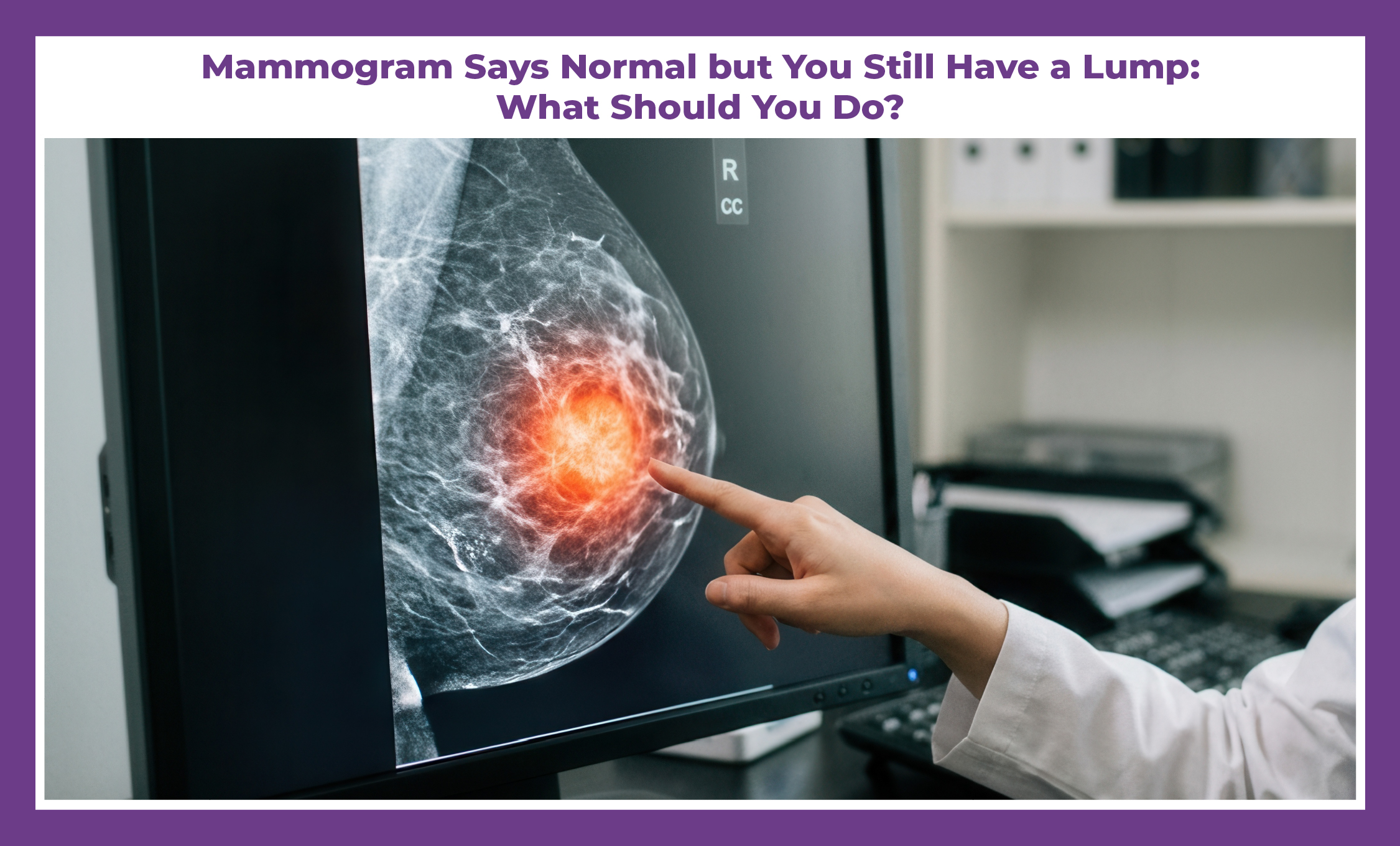
A normal mammogram is reassuring, but it does not necessarily explain every breast lump. Some lumps may be difficult to see on a mammogram, while others may be caused by benign changes that require a different type of imaging. In some situations, the physical examination and imaging may not completely agree.
That is why a persistent or new breast lump deserves proper medical evaluation, even when the mammogram report says everything is normal.
So, what should you do next?
Let's understand why this happens, what a normal mammogram actually means, when an ultrasound may be needed, and when a biopsy or specialist evaluation may be considered.
First: Don't Panic, But Don't Ignore the Lump
Feeling a lump does not automatically mean breast cancer.
Breasts naturally contain glands, ducts, fat and connective tissue. Hormonal changes can also affect how breast tissue feels throughout the menstrual cycle.
Many breast lumps turn out to be benign conditions such as cysts, fibroadenomas, fibrocystic changes or other non-cancerous abnormalities.
At the same time, a new or persistent lump should not simply be dismissed because a mammogram was normal.
The important question is:
Does the imaging actually explain the lump that you can feel?
This is sometimes referred to as clinical-imaging correlation.
Your doctor needs to consider what can be felt during examination together with what is visible on imaging.
Why Can a Mammogram Be Normal When There Is a Lump?
There isn't just one reason.
A mammogram is an important tool for detecting breast abnormalities, but like every medical test, it has limitations.
Dense breast tissue can make some abnormalities harder to see
One possible reason is dense breast tissue.
Breasts contain different proportions of fatty and glandular tissue. On a mammogram, fatty tissue generally appears darker, while dense tissue appears whiter.
Some breast abnormalities can also appear white.
As a result, abnormalities can sometimes be more difficult to distinguish from surrounding dense tissue.
his does not mean that a mammogram is useless in dense breasts. Mammography remains an important breast imaging tool. However, depending on the situation, your doctor may recommend additional imaging.
The lump may not be clearly visible on a mammogram
Not every breast lump looks the same.
Some masses may have subtle imaging features. Others may be located in an area that is technically difficult to evaluate.
This is one reason why telling your doctor exactly where you feel the lump is important.
Instead of simply saying, “I have a lump in my breast,” try to identify the location as accurately as possible.
Your doctor can then determine whether the area you are feeling corresponds to anything seen on the imaging.
The lump may be a benign cyst or another non-cancerous change
A palpable lump can have several benign causes.
For example, a breast cyst is a fluid-filled sac that can feel like a lump. Some cysts may change in size or tenderness with hormonal fluctuations.
A palpable lump can have several benign causes.
Fibroadenomas are another common benign breast condition, particularly among younger women.
Other possibilities include fibrocystic breast changes, fat necrosis, inflammation and other benign conditions.
This is why feeling a lump does not automatically mean that you have cancer.
What you feel may be normal breast tissue
Sometimes an area that feels different from the surrounding tissue may actually represent normal breast tissue.
Breasts are not perfectly uniform, and the texture can vary from one area to another.
A doctor can help determine whether what you are feeling is a distinct mass or simply an area of normal tissue.
The mammogram may not completely answer the clinical question
A mammogram provides images of the breast, but your doctor also has information that the image alone cannot provide.
For example:
- How long have you felt the lump?
- Has it become larger?
- Is it painful?
- Does it move?
- Is it firm?
- Are there changes in the skin?
- Are there nipple changes?
- Is there an enlarged lymph node?
All of these factors can influence what happens next.
That is why a normal mammogram should be interpreted alongside your symptoms and clinical examination.
What Should You Do If Your Mammogram Is Normal but the Lump Is Still There?
If you can still clearly feel the lump, the next step should generally be medical evaluation rather than simply waiting for your next routine mammogram.
Your doctor may follow several steps.
Start With a Clinical Breast Examination
A doctor will examine the breast and the surrounding area.
They may assess the lump's:
- Size
- Shape
- Location
- Texture
- Mobility
- Tenderness
They may also examine the skin, nipple and nearby lymph nodes.
The purpose is not to diagnose cancer simply by touch.
Instead, the examination helps determine whether the lump appears concerning and whether the imaging adequately explains what is being felt.
Your Doctor May Recommend a Targeted Breast Ultrasound
If you have a palpable lump and the mammogram does not provide a clear explanation, a targeted breast ultrasound may be recommended.
Unlike a mammogram, which uses X-rays to create images of the breast, ultrasound uses sound waves.
One useful feature of ultrasound is that it can help determine whether a lump is:
- Fluid-filled
- Solid
- Cystic
- Benign-appearing
- Suspicious
Ultrasound can also be used to guide a biopsy if tissue sampling becomes necessary.
The American College of Radiology includes ultrasound among the imaging tools used in the evaluation of palpable breast masses, with the appropriate approach depending on factors such as age and clinical presentation.
Is a Breast Ultrasound Better Than a Mammogram?
It is not really a question of one being universally “better.”
How Early Detection Affects Treatment Outcomes?
Mammography is particularly important for detecting certain breast abnormalities and is widely used for breast cancer screening.
Ultrasound can provide additional information about a palpable lump, particularly when the mammogram does not clearly explain what is being felt.
In some situations, both tests are used together.
The appropriate imaging depends on your age, symptoms, breast tissue and clinical findings.
What If Both Mammogram and Ultrasound Are Normal?
This is an important situation.
Suppose you have:
- A normal mammogram
- A normal ultrasound
- But you can still clearly feel a persistent lump
What happens then?
The answer depends on the clinical examination.
If the doctor believes the lump is benign and the imaging findings adequately explain the situation, monitoring may sometimes be appropriate.
But if the physical examination is concerning or the lump remains unexplained, further evaluation may be required.
The American College of Radiology emphasizes that imaging findings should be correlated with the clinical finding. If the physical examination is strongly suspicious, negative imaging should not automatically prevent further investigation.
In other words:
“The scan is normal” does not always end the conversation.
The doctor needs to determine whether the scan and the physical examination make sense together.
When Is a Biopsy Considered?
The word biopsy can immediately sound frightening.
But a biopsy does not mean that you have cancer.
A biopsy simply means that a small sample of tissue is taken so that it can be examined under a microscope.
Your doctor may recommend a biopsy if:
- The imaging shows a suspicious abnormality
- The lump feels concerning during examination
- Imaging and examination do not match
- The lump continues to persist
- The lesion changes over time
- Tissue confirmation is needed to establish a diagnosis
Depending on the location and appearance of the lesion, different types of biopsy may be considered.
For example, an ultrasound-guided core needle biopsy can be performed when the abnormality can be seen on ultrasound.
The decision depends on the individual case.
A normal mammogram is a positive and reassuring finding, but it does not mean that every possible breast abnormality has been ruled out.
This is particularly important when you have noticed a new physical change.
The National Cancer Institute advises women to tell their healthcare provider if they notice unusual breast changes, even if they recently had a normal mammogram.
The reason is simple:
Screening tests are not a substitute for investigating a new symptom.
If something feels different from your usual breast tissue, it deserves appropriate attention.
What Breast Changes Should You Pay Attention To?
A lump is one of the most commonly recognized breast changes, but it isn't the only one.
You should speak with a doctor if you notice a new or persistent:
- Breast lump: Especially if it remains present or changes over time.
- Change in breast size or shape: A new unexplained difference between the breasts should be evaluated.
- Skin dimpling or puckering: Changes in the skin over the breast may require examination.
- Nipple inversion: A newly inverted nipple should be discussed with a doctor.
- Unusual nipple discharge: Particularly spontaneous or bloody discharge.
- Lump in the armpit: An enlarged lymph node may sometimes be associated with breast conditions and should be assessed.
- Persistent focal breast changes: Any unusual change that does not settle should be discussed with a healthcare professional.
Remember, none of these signs automatically means cancer.
They simply indicate that evaluation may be appropriate.
Should You Wait for the Lump to Go Away?
Sometimes breast tissue changes with hormonal fluctuations, and some lumps may become less noticeable over time.
However, if you have identified a new, definite or persistent lump, it is better to discuss it with a doctor rather than repeatedly checking it yourself for weeks or months.
Self-monitoring can also increase anxiety because the texture of normal breast tissue can naturally vary.
A doctor can help determine whether the finding requires imaging, follow-up or further investigation.
What Is BI-RADS and Why Does It Matter?
If you've received a mammogram or breast ultrasound report, you may see the term BI-RADS.
BI-RADS stands for Breast Imaging Reporting and Data System.
It is a standardized system used by breast imaging specialists to categorize findings and communicate recommendations.
Some commonly encountered categories include:
- BI-RADS 1: Negative
- BI-RADS 2: Benign finding
- BI-RADS 3: Probably benign
- BI-RADS 4: Suspicious abnormality
- BI-RADS 5: Highly suggestive of malignancy
- BI-RADS 6: Known biopsy-proven malignancy
The category helps guide what happens next.
But remember: the report should still be considered together with your symptoms and clinical examination.
If you have a palpable lump and your report says BI-RADS 1 or 2, but the lump remains concerning or unexplained, discuss this with your doctor.
What If You Have Dense Breasts?
Dense breasts are another reason patients sometimes become confused by mammogram results.
Breast density refers to the proportion of fibroglandular tissue compared with fatty tissue.
Having dense breasts is common and is not itself a disease.
However, dense tissue can make some abnormalities more difficult to see on a mammogram.
If your report mentions dense breasts, ask your doctor whether additional imaging is appropriate for your particular situation.
The answer isn't automatically the same for everyone.
Your age, risk factors, symptoms and physical examination all matter.
Does a Lump Always Need Surgery?
No.
This is an important point.
Finding a lump does not automatically mean that you need breast surgery.
The first priority is to understand what the lump is.
Depending on the diagnosis, management could involve:
- Reassurance
- Observation
- Follow-up imaging
- Medication in selected situations
- Needle aspiration for certain cysts
- Biopsy
- Surgery when medically indicated
Surgery is generally considered only when there is an appropriate reason for it.
This is why it is important not to jump directly from “I feel a lump” to “I need breast cancer surgery.”
Diagnosis comes first.
When Should You See a Breast Specialist?
You don't have to wait until a lump becomes painful or large before seeking medical advice.
Consider speaking with a breast specialist if:
- The lump is new
- It persists
- It is increasing in size
- You notice associated skin or nipple changes
- You have a family history that concerns you
- You have a family history that concerns you
- Different tests provide conflicting information
- You remain concerned despite a normal mammogram
A breast specialist can review your clinical examination and imaging together and determine whether additional evaluation is necessary.
For someone searching online for a Breast Cancer Doctor in Delhi, Breast Cancer Doctor in Gurgaon/Gurugram, or Breast Cancer Doctor in Haryana, the key consideration should be finding a doctor who can properly evaluate the symptom rather than simply relying on one test result.
Similarly, patients searching for Dr. Deepak Jha, a Breast Cancer Surgeon in Haryana, Best Breast Cancer Surgeon in Delhi, or Best Breast Cancer Surgeon in Gurgaon/Gurugram should understand that specialist consultation is not synonymous with surgery.
A consultation can simply be the next step in understanding what a breast lump means and whether further testing is necessary.
What Should You Take to Your Appointment?
If you are seeing a doctor about a persistent lump, take your previous reports with you.
These may include:
- Mammogram report
- Mammogram images/CD or digital access, if available
- Ultrasound reports
- Previous breast imaging
- Biopsy reports, if applicable
- Relevant medical records
Also make a note of:
When did you first notice the lump?
Has it changed in size?
Is it painful?
Does it change during your menstrual cycle?
Have you noticed skin or nipple changes?
Have you noticed any lump in the armpit?
These details can help your doctor understand the situation.
A Simple Way to Think About It
If you remember only one thing from this article, remember this:
A mammogram tells you what the imaging shows. Your hands tell you what you are feeling. Your doctor needs to put both pieces of information together.
A normal mammogram is reassuring.
“Is my mammogram normal?”
It should be:
“Has the lump I can feel been properly explained?”
If the answer is yes, your doctor can guide you on whether reassurance or follow-up is appropriate.
If the answer is no, additional evaluation may be needed.
Mammogram Normal but Lump Still There: What Should You Do?
Don't panic.
Don't assume the lump is cancer.
But don't ignore it either.
Start with a clinical examination. Depending on your age and individual situation, your doctor may recommend targeted ultrasound or other imaging. If the clinical examination and imaging do not agree, further investigation, including biopsy in selected cases, may be considered.
Most importantly, do not let a normal mammogram prevent you from reporting a persistent breast change.
Breast health is not about being frightened by every lump. It is about recognizing changes, getting the right evaluation and understanding what your results actually mean.
If you are looking for a Breast Cancer Doctor in Delhi, Breast Cancer Doctor in Gurgaon/Gurugram, Breast Cancer Doctor in Haryana, or a specialist such as Dr. Deepak Jha, discussing a persistent breast lump with an experienced breast specialist can help you understand the appropriate next step.
And if further treatment is eventually required, options such as Breast Cancer Surgery in Delhi, Breast Cancer Surgery in Gurgaon/Gurugram, or Breast Cancer Surgery in Haryana can be discussed only after the diagnosis and treatment plan are established.
A normal report is good news. But a persistent lump still deserves an answer.
This article is intended for general educational purposes and does not replace an in-person medical consultation, examination or individualized medical advice.
Subscribe Newsletter
Get all the information, latest updates, and articles delivered directly in your inbox